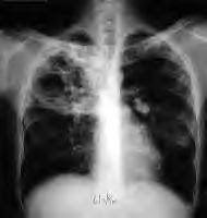
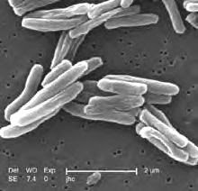
Smear examination for the detection of Acid fast bacilli continues to be the gold standard in Diagnosis of Tuberculosis. In spite of several inadequacies, Microscopy for AFB detection is economical, specific and the man power can be trained easily. The detection of AFB in sputum smears helps for higher case detection, and contains the spread of Tuberculosis in the Society.
The smear will become Postive when one has bacilli more than 5,000 – 10, 000 / 1 ml of sputum. Multiple smear examinations, at least three morning specimens are advised and appropriate collection of specimens will increase yield to > 43 %. If efforts were taken in educating patients for 1- 2 minutes in methods to collect the sputum, will yield higher results. Sputum induction procedures are helpful. Today’s emphasis to identify AFB, in smears is more demanding with associated HIV/AIDS, as few bacilli are excreted. Concentration of specimens and digestion of thick and mucous associate specimens with Sodium hypochlorite, Sodium hydroxide, N-acetyl –cystine – Sodium hydroxide will increase rate of detection to > 18 % in sensitivity and incremental yield of 9 %( positve after treatment with above chemicals – positives with direct Ziehl Nelson’s straining ) Sodium hypochlorite is beneficial in HIV positive patients as it is Mycobactericidal and also kills human Immunodeficiency virus, but not suitable for culturing specimens.
Need for Florescent Microscopy
The developing world should explore the Fluorescence microscopy, which will improve the sensitivity of Microscopy in patient who excrete few bacilli as in association HIV infection, The role of Ziehl Neelsen’s method of staining and conventional Microcopy is losing the sensitivity with ever increasing work load, technicians opting to see few fields, monotonous nature of work, the lack of accountability, and inter Institutional quality control protocols. Many systematic reviews indicated use of Florescent Microscopy will increase 10% higher sensitivity and 9 % in incremental yield when compared with Z.N method of staining. About 15 times as many fields of view can be scanned by Fluorescent Microscopy as by conventional Microscopy in the same period. The developing countries face crunch to buy Fluorescent microscopes and to maintain the regular availability of florescent dyes. It is utmost important to develop centralized and dedicated centers for Microscopy to have control on peripheral laboratories. Negative smears by conventional Microscopy needs further attention with optimal microscopy, concentration methods to detect AFB to reduce early mortality among the infected and to contain the spread in society.
Culturing for Mycobacterial Isolation
Sputum culturing remains a gold standard for diagnosing Mycobacterial infections. A Postive grwoth can be demonstrated with few bacilli to as low as 10 – 100 of viable bacilli per I ml of sputum. Cultures show growth of AFB even when patients where on treatment and negative by smear examination. A simple measure with decontamination of specimens and inoculation of at least 150 – 200 µl of concentrate on culture medium will increase the success in culturing. In spite of best decontamination procedures, 1 – 4 % of the isolates are false Postive. The greatest limitation of culturing on Lowenstein – Jensen medium and other equivalent medium is long periods (2 – 12 weeks) for isolation of bacteria.
Advances in Diagnostic Methodologies.
1. Mycobacterial growth in Incubator tube MGIT (Mycobacterium Growth Indicator Tube) is one new culturing method, costlier to install and automated system. Economic limitations and timely availability of reagents (closed system committed to the manufactures.) continue to hamper the growth of technolology in developing world
2.. Recent success with MODS ( the Microscopic Observation of drug susceptibility Assay ) developed in Peru gained the success as affordable, and primary drug resistance can be performed with simple efforts, But inverted microscope is essential to read the results at frequent intervals. Contamination or hazard to technical personnel is minimal. Even the district laboratories can report resistance to Isoniazid and Rifampicin In spite of several controlled studies on MODS assay is poor to discriminate between, M.tuberculosis from Non Tuberculosis Mycobacterium. The success of MODS is a great breakthrough in detection of MDR strains provided the prevalence of NTM prevalence is low MODS assay can identify patients with TB in approximately on third of time required for culturing on L J medium.
Emerging and Rapid Diagnostic methods.-
1 Fast plaque with phage amplification technology, tested in areas with high rates of HIV infection, had contradicting results, needs more understanding.
2. Quanti – Feron TB test – Done on Blood specimens, based on the principle of ELISA and enzyme linked immunospot. With higher production of Interferon γ (Inf-γ) by cells in response to Mycobacterium tuberculosis, than to the other environmental Mycobacterium in particular to Mycobacterium avium complex. The testing results correlated with Tuberculin skin test reactivity, but still hampered in BCG vaccinated.
3 Elispot – Tested by Elisa methodology detects Interferon γ produced by T lymphocytes in response to latent Tuberculosis
Infection. Elispot gained more clinical acceptability and advantageous, being negative in majority of BCG vaccinated individuals
Both the above testing methods were limited to high end laboratories and cost of testing remained the major limitation in many developing countries. More helpful to diagnose the latent Tuberculosis Infections.
Molecular Technology:
The fast gains of Polymerase technologies by amplification of DNA (PCR) are limited to controlled studies interpreted in relation to clinical context and performance of the laboratory .Rapidly changing molecular technologies, out dating earlier hardware, other equipment and patented primers, added to limitations in the Developing world. Mainly used as restricted research tool, and unaffordable to the needy poor.
Many extra pulmonary tuberculosis cases were benefited with Molecular technology.
Future Goals in Control of Tuberculosis ;
Stop TB partnership, Global Plan for 2006 to 2015 call for strengthening of network to facilitate detect all TB cases including smear negative tuberculosis. The Emphasis should focus on Sputum concentration methods, promoting the use of Fluorescent Microscopy. Helping the smaller laboratories to initiate culturing, and antibiotic sensitivity testing. The present affordable option may remain with utilizing the methodology of MODS .The Developing world wishes to utilize this upcoming technology for practical, and simple way to detect the MDR tuberculosis even at district Laboratories
. Yet there is no fool proof, sensitive and specific test, which is inexpensive and rapid method for Diagnosing the Tuberculosis.
Great challenges include detection and controlling of MDR TB. Strengthening the Smear Microscopy, and more aggressive provisions for enforcing the Fluorescent microscopy, may reduce the incidence of spread of tuberculosis. We have to watch the Impact of X-MDR in the Indian continent. The undergraduate and postgraduate Medical students should be taught with more emphasis on control of drug resistant tuberculosis The best options with implementation of International standards for tuberculosis care with initiation of Major global health participation may bring hope to reduce the incidence of Tuberculosis by 2015.
http://fieldmethods.com/?p=127
Subscribe to:
Post Comments (Atom)


No comments:
Post a Comment